Tumor Spheroid - Real volume of spheroids

Real volume of spheroids

Objective

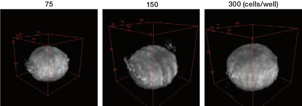
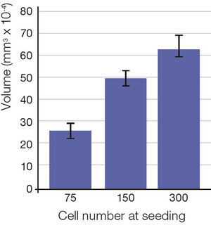
The volume of spheroids that stepwise cell number seeding, is quantified by using Cell3iMager Estier.

Tumor Spheroid - Real volume of spheroids  - Spheroids tomographic image

Materials and Methods


Cell Line

MCF7 cells (RIKEN BRC)

Medium

DMEM (Nacalai tesque)

Plate

96-well plate U bottom (Sumitomo Bakelite)

Seeding cell density

75 - 300 cells/well

Culture days

3 days

Imaging methods

Low magnification lens
Each scan 3 um pitch

Image processing and analysis (ImageJ)

Median 3D filter (X, Y, Z radius; 2.0, 2.0, 2.0)
3D Object Counter v2.0(Threshold; 8000)

Results and Conclusions

  • It is possible to get the three-dimensional image from label-free samples by using Cell3iMager Estier.
  • It is showed that the volume of spheroids depend on stepwise cell number seeding.
  • It is possible to quantify the exact volume of spheroids that was difficult before.
  • Cell3iMager Estier Estier can observe the internal structure of spheroids from tomograms.

It is possible to quantify the exact volume of spheroids that was difficult before.                           It is possible to quantify the exact volume of spheroids that was difficult before.

Related Product

Would you like additional information?

I agree to the terms and conditions

I agree to the GDPR

Need help finding the right product?
Get in touch
+1 (844) 454-3399